
Common Symptoms
- Lateral elbow pain that is sharp with attempted forearm rotation (pronation and supination)
- Tenderness directly over the radial head, palpable just distal to the lateral epicondyle
- Swelling in the lateral elbow with a positive posterior fat pad sign on radiograph
- Limited forearm rotation, especially supination, due to pain and mechanical block
- Painful arc during elbow flexion and extension when the fracture fragment impinges
- Crepitus with passive forearm rotation in comminuted or displaced fractures
Radial Head Fracture Imaging Signs: Fat Pad Sign and Mason Classification
Your elbow X-ray shows a radial head fracture? Learn what Mason types 1 to 4 mean, why a fat pad sign matters and which findings can change treatment.
Overview
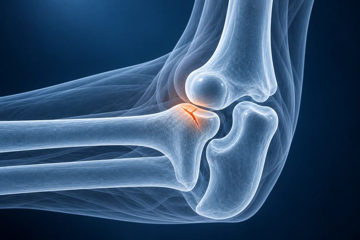
Radial head fractures are the most common elbow fracture in adults, typically resulting from a fall onto an outstretched hand. The Mason classification guides treatment decisions. Our AI consortium evaluates fracture pattern, displacement, comminution, and associated injuries including the terrible triad (radial head fracture + coronoid fracture + elbow dislocation).
Key Imaging Findings
- Fracture line through the radial head or neck
- Mason classification: Type I (non-displaced), II (displaced), III (comminuted)
- Anterior or posterior fat pad sign on lateral X-ray
- Associated coronoid process fracture
- MCL or LCL injury on MRI
- Occult fracture visible on MRI but not X-ray
Frequently Asked Questions
What is the Mason classification of radial head fractures?
The Mason classification categorizes radial head fractures into four types. Type I fractures are non-displaced or minimally displaced (less than 2 mm) with no mechanical block; these are treated non-operatively with early mobilization. Type II fractures involve displacement of greater than 2 mm or greater than 30 degrees of angulation and may require fixation if a mechanical block to forearm rotation exists. Type III fractures are comminuted involving the entire radial head, typically requiring radial head replacement rather than fixation. Type IV fractures, added by Johnston, are any radial head fracture associated with an elbow dislocation, significantly increasing instability and complexity of management.
How is a radial head fracture diagnosed when plain radiographs appear normal?
A posterior (sail sign) fat pad on lateral elbow radiograph in the setting of trauma indicates a joint effusion and a presumed occult fracture, most commonly a radial head fracture, even when the fracture line itself is not visible. The anterior fat pad, when elevated and sail-shaped rather than lying flat, also supports an effusion. If radiographs are equivocal and a fracture is suspected clinically, CT provides superior fracture characterization including fragment size, displacement, and comminution, and is essential for surgical planning. MRI is useful when associated ligament injuries or chondral damage need assessment.
Can a radial head fracture be associated with other injuries?
Yes. Radial head fractures frequently occur alongside other elbow and forearm injuries. The terrible triad injury combines elbow dislocation, radial head fracture, and coronoid fracture, creating multiplanar instability requiring comprehensive surgical reconstruction. An Essex-Lopresti injury involves a radial head fracture with disruption of the interosseous membrane and distal radioulnar joint, leading to proximal radial migration if the radial head is excised without replacement. Medial collateral ligament tears often accompany radial head fractures and must be assessed under fluoroscopic valgus stress testing.
Sources
- Mason ML. Some observations on fractures of the head of the radius with a review of one hundred cases. Br J Surg. 1954;42(172):123-132.
- Johnston GW. A follow-up of one hundred cases of fracture of the head of the radius with a review of the literature. Ulster Med J. 1962;31(1):51-56.
- Hotchkiss RN. Displaced fractures of the radial head: internal fixation or excision? J Am Acad Orthop Surg. 1997;5(1):1-10.
- Goswami GK. The fat pad sign. Radiology. 2002;222(2):419-420.
Get an educational explanation of imaging terms related to Radial Head Fracture
Upload your MRI or X-ray DICOM files for private, AI-powered analysis. 4 models analyze independently — raw scan files stay in your browser.
Upload & AnalyzeMedical Disclaimer: This page is for informational and educational purposes only. It does not constitute medical advice, diagnosis, or treatment. AI-generated analysis may contain errors. Always consult a qualified healthcare professional for medical decisions. Full Disclaimer