
MRI vs Ultrasound for Elbow Injuries: When to Use Each
Compare MRI and ultrasound for tennis elbow, golfer's elbow, UCL tears, and biceps injuries — strengths, limitations, cost, and dynamic assessment.
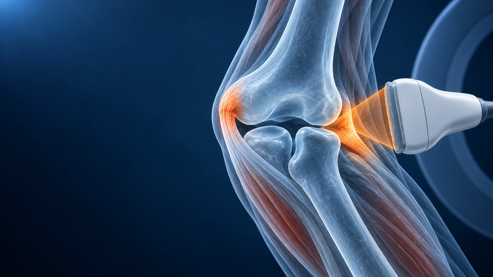
Both MRI and musculoskeletal ultrasound are valuable tools for evaluating elbow injuries, and in many clinical situations your physician will choose between them based on the suspected diagnosis, available equipment, and practical considerations such as cost and urgency. Neither modality uses ionizing radiation, so there is no X-ray exposure with either test.
Ultrasound excels at dynamic assessment — the ability to move the joint during imaging — and is faster, less expensive, and available at the point of care. MRI provides a comprehensive single-snapshot of all structures simultaneously, including deep bone marrow, cartilage, and intra-articular pathology that ultrasound cannot reliably reach. Understanding the strengths and limitations of each helps you have a more informed conversation with your orthopedic surgeon or sports medicine physician.
When MRI Wins
MRI is the preferred modality when deep structures or bone involvement is suspected. It is the gold standard for evaluating osteochondritis dissecans (OCD) of the capitellum, where subchondral bone signal changes and overlying cartilage integrity must be assessed to stage the lesion and decide between conservative and surgical treatment. MRI is also superior for detecting bone marrow edema from stress reactions, occult fractures not visible on X-ray, and intra-articular loose bodies when contrast arthrography is used.
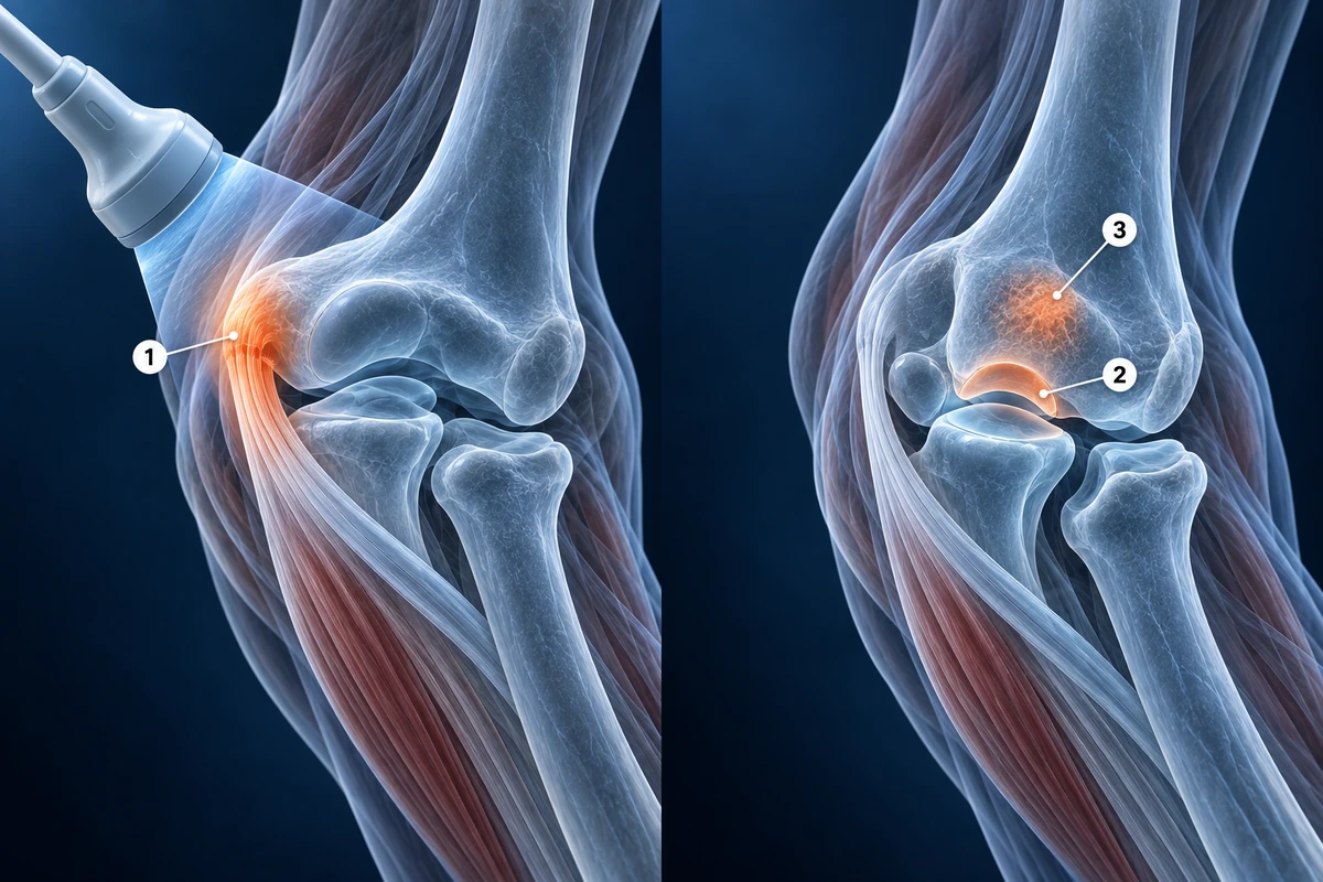
For comprehensive single-session assessment — when a clinician wants to evaluate ligaments, tendons, nerves, cartilage, and bone simultaneously — MRI is unmatched. MR arthrography (with intra-articular gadolinium injection) significantly improves sensitivity for partial UCL tears and cartilage defects that may be occult on standard sequences. See our guide on how to read elbow MRI for a detailed walkthrough of sequences and findings.
When Ultrasound Wins
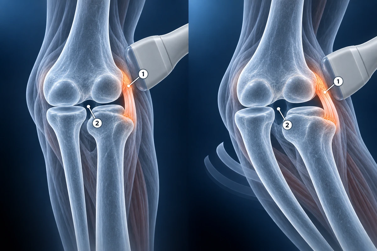
Ultrasound has a decisive advantage when dynamic stress testing is needed. Applying valgus stress to the medial elbow during real-time imaging allows direct visualization of UCL laxity and joint space widening — a finding that static MRI cannot replicate. This dynamic assessment is particularly valuable in throwing athletes where subtle UCL insufficiency may only manifest under load. Ultrasound is also preferred for serial monitoring of common extensor tendon healing in lateral epicondylitis, where changes in tendon echogenicity and thickness can be tracked over treatment.
Practical advantages favor ultrasound in many clinical settings: a typical musculoskeletal ultrasound costs approximately $150–$400 compared to $1,000–$3,000 for MRI without insurance. Ultrasound is available at the point of care in many sports medicine clinics, requires no patient preparation, and can guide real-time injection therapy — corticosteroid, platelet-rich plasma, or barbotage for calcific tendinopathy — with direct needle visualization. The primary limitations are operator dependence and reduced penetration in patients with a larger body habitus, where deep structures may be obscured.
Tennis Elbow (Lateral Epicondylitis)
Both modalities are effective for lateral epicondylitis. On ultrasound, the common extensor tendon origin appears hypoechoic (darker than normal) with tendon thickening, loss of the normal fibrillar echotexture, and occasionally intratendinous calcifications that cast an acoustic shadow. Neovascularity on power Doppler indicates active tendinopathy. Ultrasound sensitivity for full-thickness common extensor tears approaches 80–95% and specificity exceeds 90%.
On MRI, lateral epicondylitis presents as increased T2 signal within the common extensor tendon, tendon thickening, and peritendinous edema. Tendinosis without tearing shows intermediate signal on T2; partial tearing introduces fluid-signal intensity within the substance; complete tears show full discontinuity. MRI sensitivity for significant tendinopathy approaches 90–100% and additionally provides information about the radial collateral ligament and posterolateral rotatory stability that ultrasound cannot assess reliably. For a deeper look at lateral epicondylitis, visit the lateral epicondylitis condition page.
Golfer's Elbow (Medial Epicondylitis)
Medial epicondylitis follows similar imaging parity between modalities. On ultrasound, the flexor-pronator tendon origin shows hypoechogenicity, thickening, and Doppler flow comparable to what is seen at the lateral epicondyle in tennis elbow. MRI shows increased T2 signal in the common flexor tendon with associated bone marrow edema at the medial epicondyle in more severe cases. Because the ulnar nerve runs in close proximity to the medial epicondyle, both modalities should assess for concurrent cubital tunnel pathology, though MRI provides more reliable nerve cross-sectional area measurement and surrounding edema characterization.
UCL Tears
Ultrasound dynamic valgus stress testing is uniquely valuable for UCL assessment in throwing athletes. Joint space widening of more than 1 mm under valgus load compared to the contralateral elbow is considered a positive finding for UCL insufficiency. This real-time functional assessment complements the structural information obtained from cross-sectional imaging. Ultrasound sensitivity for complete UCL tears is approximately 72–88% in experienced hands, rising with dynamic stress.
MRI and MR arthrography remain the gold standard for full characterization of UCL pathology. Standard MRI identifies complete tears with high accuracy, but partial undersurface tears — the most clinically significant injury pattern in overhead athletes — may be missed without intra-articular gadolinium. MR arthrography increases sensitivity for partial tears to approximately 86–92% by allowing contrast to track through the tear site (the T-sign: contrast pooling between the UCL and the sublime tubercle on coronal images). When surgical planning is required, MR arthrography is the preferred pre-operative study.
Distal Biceps Rupture
Ultrasound is an excellent rapid first-line assessment for suspected distal biceps rupture. Dynamic maneuvers — asking the patient to supinate the forearm while scanning — can demonstrate the retracted tendon end and the fluid-filled empty bicipital tunnel in complete tears. Ultrasound can be performed immediately in the emergency or office setting, before swelling and pain limit range of motion. Sensitivity for complete distal biceps tears on ultrasound exceeds 95% in experienced hands.
MRI provides definitive characterization for surgical planning, particularly in partial tears where the degree of fiber involvement determines operative versus conservative management. The FABS (flexed elbow, abducted shoulder, supinated forearm) MRI position allows the distal biceps tendon to be visualized along its full length on a single coronal image, significantly improving assessment of the footprint at the radial tuberosity. MRI is also superior for evaluating associated injuries to the bicipital bursa and surrounding structures.
Key Takeaways
- Neither MRI nor ultrasound uses ionizing radiation — both are radiation-free modalities
- Ultrasound dynamic valgus stress testing is unique to ultrasound and is valuable for UCL insufficiency in throwing athletes
- MRI with arthrography is the gold standard for partial UCL tears and pre-operative surgical planning
- Ultrasound costs approximately $150–$400 versus $1,000–$3,000 for MRI, and allows real-time injection guidance
- OCD of the capitellum and bone marrow edema require MRI — ultrasound cannot reliably assess these deep structures
- Ultrasound accuracy depends heavily on operator experience; MRI provides a reproducible permanent record readable by any radiologist
Frequently Asked Questions
Which is cheaper — elbow MRI or elbow ultrasound?
Ultrasound is substantially less expensive. A musculoskeletal elbow ultrasound typically costs $150–$400 out of pocket, while MRI ranges from $1,000–$3,000 without insurance depending on facility and whether contrast is used. Insurance coverage varies and often requires prior authorization for MRI. In many sports medicine clinics, ultrasound can be performed in the same appointment as the consultation, with no scheduling delay.
Does elbow MRI or ultrasound involve radiation?
Neither modality involves ionizing radiation. MRI uses magnetic fields and radio waves; ultrasound uses high-frequency sound waves. Both are considered safe for repeated use, which is particularly relevant when monitoring recovery from tendinopathy or following a partial tear conservatively. X-ray and CT do involve radiation and are used for different indications — primarily bony pathology.
Can ultrasound see a UCL tear fully?
Ultrasound can identify complete UCL tears with reasonable accuracy (approximately 72–88% sensitivity in experienced hands) and adds the unique ability to perform dynamic valgus stress testing. However, partial undersurface tears — the most common pattern in overhead throwing athletes — are frequently missed on ultrasound alone. MR arthrography with intra-articular contrast is the most sensitive test for partial UCL tears and is preferred when surgical decision-making is required.
Do I need contrast for an elbow MRI?
Standard MRI without contrast is sufficient for most elbow conditions including epicondylitis, distal biceps assessment, nerve evaluation, and most tendon injuries. MR arthrography — where gadolinium contrast is injected directly into the joint before imaging — is specifically recommended when evaluating partial UCL tears, subtle cartilage defects, loose bodies, or OCD lesion stability. The injection is performed under fluoroscopic or ultrasound guidance and adds approximately 30 minutes to the procedure.
How much does operator skill affect ultrasound accuracy?
Operator experience has a significant impact on ultrasound accuracy — more so than with MRI, where the images are recorded and can be re-reviewed by any radiologist. Published sensitivity figures for musculoskeletal ultrasound reflect results from experienced operators at specialist centers; accuracy at general facilities may be lower. When seeking ultrasound for elbow pathology, a radiologist or sports medicine physician with specific musculoskeletal ultrasound training will produce more reliable results than a general ultrasound technologist.
Related Articles
Understand your elbow MRI report including UCL evaluation, epicondylitis findings, and OCD lesion staging.
Understand common elbow conditions including tennis elbow, golfer's elbow, UCL tears, radial head fractures, and OCD lesions.
Related Conditions
Ready to analyze your imaging? Upload your MRI or X-ray for AI-powered analysis
Upload your MRI or X-ray DICOM files for private, AI-powered analysis. 4 models analyze independently — raw scan files stay in your browser.
Start AnalysisMedical Disclaimer: This page is for informational and educational purposes only. It does not constitute medical advice, diagnosis, or treatment. AI-generated analysis may contain errors. Always consult a qualified healthcare professional for medical decisions. Full Disclaimer