
Elbow Dislocation on MRI & X-ray: Imaging Findings
The elbow is the second most commonly dislocated major joint in adults. Associated fractures and ligament injuries need careful assessment, and rehabilitation depends on stability after reduction.
Some signs described on this page need prompt medical care. If you have them, get care first. Reading about imaging can wait.
Common Symptoms
- Severe pain immediately after a fall on an outstretched hand with the elbow partially flexed
- Visible gross deformity of the elbow with the olecranon prominent posteriorly
- Inability to move the elbow actively and guarding against any passive manipulation
- Rapid and diffuse swelling that obscures normal bony landmarks within minutes of injury
- Numbness or paresthesias in the median or ulnar nerve distribution if traction neuropraxia has occurred
- Diminished radial pulse or pale, cool hand indicating vascular compromise requiring emergency evaluation
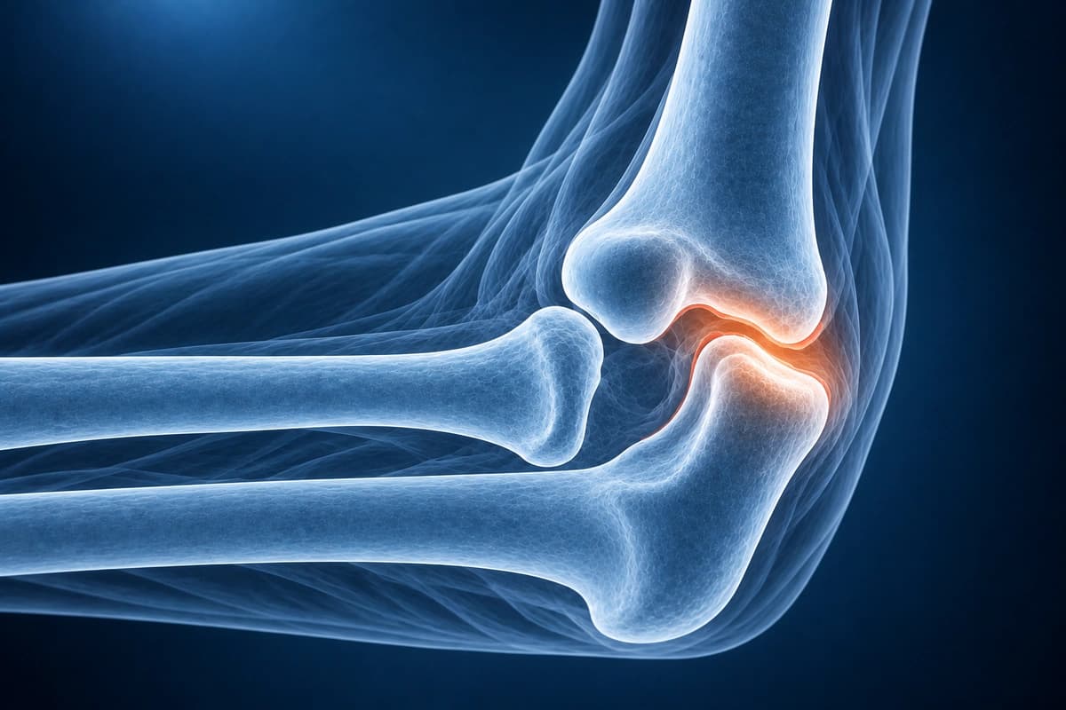
What We Look For on Imaging
- Joint congruity assessment on post-reduction imaging
- Coronoid process fracture type and fragment size (Regan-Morrey classification)
- Radial head fracture and degree of comminution
- Lateral ulnar collateral ligament tear indicating posterolateral rotatory instability
- Medial collateral ligament disruption and extent of tearing
- Intra-articular loose bodies and capsular disruption
Frequently Asked Questions
What structures are injured in a simple versus complex elbow dislocation?
Simple elbow dislocations involve purely capsuloligamentous injury without associated fractures. The lateral ulnar collateral ligament is the primary restraint disrupted, followed sequentially by the anterior capsule and the medial ulnar collateral ligament in the Horii circle of disruption described by O'Driscoll. Complex elbow dislocations involve associated fractures of the radial head, coronoid, or both, constituting the terrible triad injury. Coronoid fractures are classified by the Regan–Morrey system (Type I: tip avulsion, Type II: up to 50% of coronoid, Type III: greater than 50%) or the O'Driscoll classification, with larger fragments representing greater instability.
How should an elbow dislocation be reduced and when is urgent reduction required?
Closed reduction should be performed as soon as possible, ideally in the emergency department with adequate analgesia and procedural sedation. For a posterior dislocation, the most common direction, traction is applied to the forearm along its long axis while the elbow is gently flexed, and the olecranon is simultaneously lifted anteriorly. Neurovascular examination before and after reduction is mandatory. Vascular compromise—absent radial pulse, hand pallor, or expanding hematoma—is an indication for emergency reduction, and if the pulse does not return after reduction, vascular surgery consultation for arterial exploration is required, as the brachial artery is at risk in severely displaced posterolateral dislocations.
What is the expected recovery timeline after elbow dislocation?
After successful closed reduction of a simple elbow dislocation, the joint is splinted in 90 degrees of flexion for 7 to 10 days to allow initial soft tissue healing, followed by progressive range-of-motion exercises. Most patients regain functional motion within 6 to 8 weeks, though some residual extension loss of 10 to 15 degrees is common. Return to sport occurs at 3 to 6 months. Complex dislocation with fractures typically requires surgical fixation of the radial head and coronoid, ligament repair or reconstruction, and a longer rehabilitation course of 6 to 12 months. Stiffness is the most common long-term complication, and heterotopic ossification can develop in high-energy injuries.
Sources
- Kuhn MA, Ross G. Acute elbow dislocations. Orthop Clin North Am. 2008;39(2):155-161.
- O'Driscoll SW, Morrey BF, Korinek S, An KN. Elbow subluxation and dislocation. A spectrum of instability. Clin Orthop Relat Res. 1992;(280):186-197.
- Regan W, Morrey B. Fractures of the coronoid process of the ulna. J Bone Joint Surg Am. 1989;71(9):1348-1354.
- Ring D, Jupiter JB, Zilberfarb J. Posterior dislocation of the elbow with fractures of the radial head and coronoid. J Bone Joint Surg Am. 2002;84(4):547-551.
- Mehlhoff TL, Noble PC, Bennett JB, Tullos HS. Simple dislocation of the elbow in the adult. Results after closed treatment. J Bone Joint Surg Am. 1988;70(2):244-249.
Do not delay urgent medical care to use this tool.
After you get care, understand your imaging
MRI, X-ray or CT from a CD, folder or ZIP. A PDF report or photos of the films work too.
Free to open and preview, no account needed. An optional AI report from $10 explains your images in plain language, with questions for your doctor.
Your raw files stay on this device. Only rendered images and minimal context are sent when you request an AI report.
Medical Disclaimer: This page is for informational and educational purposes only. It does not constitute medical advice, diagnosis, or treatment. AI-generated analysis may contain errors. Always consult a qualified healthcare professional for medical decisions. Full Disclaimer