
Elbow Arthritis on X-ray: Imaging Findings
Elbow arthritis can be primary (rare), post-traumatic, or inflammatory (rheumatoid). Post-traumatic arthritis is the most common form, developing after fractures or dislocations.
Common Symptoms
- Gradually worsening aching or deep pain in the elbow that increases with activity and at end range of motion
- Mechanical loss of terminal extension and flexion due to osteophytes and capsular contracture
- Locking or catching sensation caused by loose osteocartilaginous bodies within the joint
- Crepitus during elbow flexion and extension that may be audible as well as palpable
- Morning stiffness lasting less than 30 minutes in primary osteoarthritis, longer in inflammatory arthritis
- Ulnar nerve symptoms including ring and little finger tingling from nerve tethering by medial osteophytes
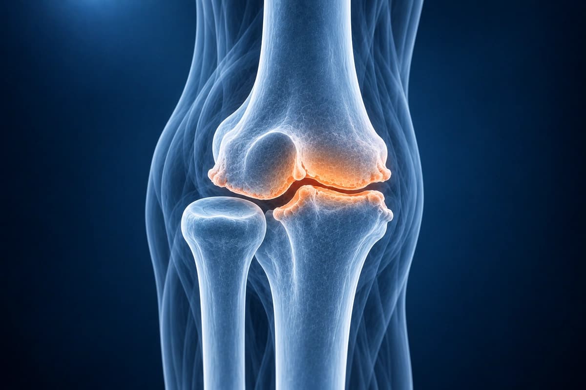
Key Imaging Findings
- Joint space narrowing on X-ray
- Olecranon and coronoid osteophytes
- Loose bodies within the joint
- Olecranon fossa osteophytes causing impingement
- Cartilage thinning and defects on MRI
- Synovitis and joint effusion
Frequently Asked Questions
Who is most commonly affected by primary elbow osteoarthritis?
Primary elbow osteoarthritis disproportionately affects middle-aged male manual laborers and overhead athletes with a history of heavy repetitive use, in contrast to the age- and female-predominant pattern seen in knee and hip osteoarthritis. The dominant extremity is involved in most cases. Radiographs show posterior olecranon and coronoid osteophytes, olecranon fossa impingement, and loose bodies, with relative preservation of joint space in early disease distinguishing it from inflammatory arthritis where global joint space narrowing occurs early.
How does rheumatoid arthritis of the elbow differ from osteoarthritis?
Rheumatoid arthritis causes synovial proliferation that erodes articular cartilage globally, leading to concentric joint space loss, periarticular osteopenia, and marginal erosions on radiograph. Inflammatory markers (ESR, CRP, rheumatoid factor, anti-CCP antibodies) are typically elevated. Patients experience prolonged morning stiffness exceeding one hour and systemic features such as fatigue and involvement of multiple small joints. In contrast, primary elbow osteoarthritis shows preserved or minimal joint space loss early, marginal osteophytes, and normal inflammatory markers, with pain triggered by mechanical loading rather than rest.
When is total elbow replacement considered for elbow arthritis?
Total elbow arthroplasty is most appropriate for elderly, low-demand patients with inflammatory arthritis (particularly rheumatoid) who have failed medical management, or for severe post-traumatic arthritis with global joint destruction in older individuals. The semi-constrained linked design is the most widely used implant. Because the elbow experiences high cyclic loading relative to its small contact area, weight-bearing restrictions of 5 to 10 pounds for lifting are imposed for life, making total elbow replacement less suitable for young or active patients. For younger, higher-demand patients, arthroscopic or open debridement with osteophyte removal and capsular release provides durable symptom relief while preserving the option for future arthroplasty.
Sources
- Gramstad GD, Galatz LM. Management of elbow osteoarthritis. J Bone Joint Surg Am. 2006;88(2):421-430.
- Cheung EV, Adams R, Morrey BF. Primary osteoarthritis of the elbow: current treatment options. J Am Acad Orthop Surg. 2008;16(2):77-87.
- Papatheodorou LK, Baratz ME, Sotereanos DG. Elbow arthritis: current concepts. J Hand Surg Am. 2013;38(3):605-613.
- Antuña SA, Morrey BF, Adams RA, O'Driscoll SW. Ulnohumeral arthroplasty for primary degenerative arthritis of the elbow: long-term outcome and complications. J Bone Joint Surg Am. 2002;84(12):2168-2173.
Understand your imaging before your next appointment
MRI, X-ray or CT from a CD, folder or ZIP. A PDF report or photos of the films work too.
Free to open and preview, no account needed. An optional AI report from $10 explains your images in plain language, with questions for your doctor.
Your raw files stay on this device. Only rendered images and minimal context are sent when you request an AI report.
Medical Disclaimer: This page is for informational and educational purposes only. It does not constitute medical advice, diagnosis, or treatment. AI-generated analysis may contain errors. Always consult a qualified healthcare professional for medical decisions. Full Disclaimer