Elbow Fracture Imaging Signs: Fat Pad Effusion, Alignment, and Displacement
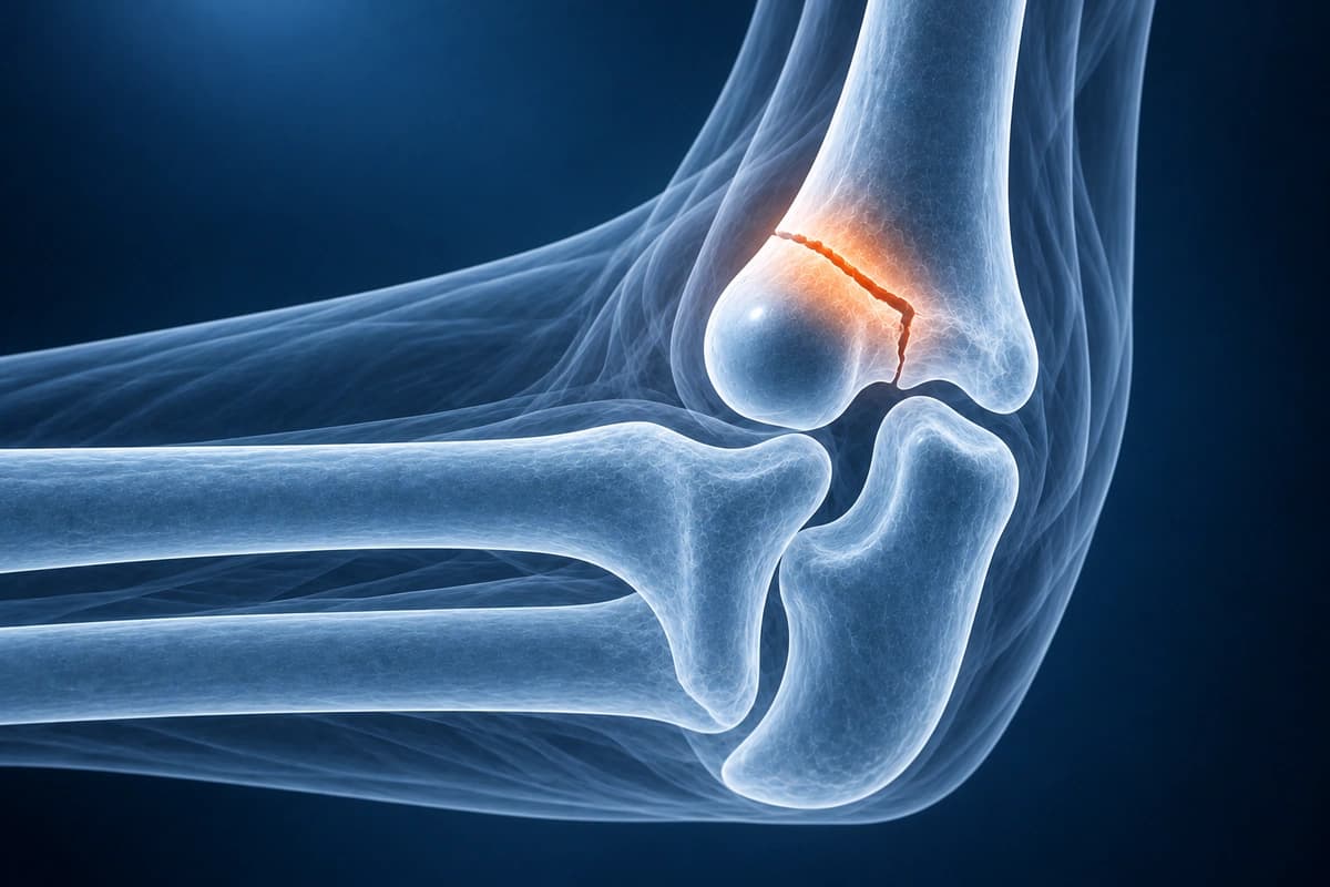
A “positive fat pad sign” means the side-view elbow X-ray shows the fat pads lifted away from the bone by fluid inside the joint, called an effusion. After an injury, that effusion often comes with a fracture too small to see on the X-ray. It is a warning sign, not a fracture diagnosis on its own.
Common Symptoms
- Immediate and severe pain at the elbow following a fall, direct blow, or high-energy trauma
- Visible deformity, shortening, or angular malalignment of the elbow joint
- Rapid swelling and ecchymosis spreading across the antecubital fossa and distal arm
- Inability to actively extend or flex the elbow due to pain and mechanical block
- Neurovascular compromise including wrist drop, weak grip, or diminished radial pulse
- Crepitus on gentle palpation indicating disrupted bone cortex
Fat Pad Sign on Your Elbow X-ray Report
How the finding is usually worded
- “Positive anterior and posterior fat pad sign”
- “Elevated anterior fat pad” or “sail sign”
- “Visible posterior fat pad”
- “Elbow joint effusion, no displaced fracture identified”
- “Effusion, suspect occult radial head fracture”
Anterior versus posterior fat pad
On a true lateral view with the elbow bent to 90 degrees, a thin anterior fat pad normally lies flat against the front of the humerus, and the posterior fat pad stays hidden in its bony hollow. Fluid in the joint lifts the anterior pad into a triangle, the sail sign, and pushes the posterior pad into view. A visible posterior fat pad is considered abnormal.
How often a hidden fracture is really there differs between studies. In adults with an effusion and no visible fracture, MRI found an occult fracture in 75%, most often of the radial head, and the size of the effusion did not predict which elbows were broken. In children, follow-up X-rays showed a healing fracture in 76% in one study but only 17% in another.
Why it changes the next steps
Because a fracture is often present, many clinicians manage an injured elbow with a fat pad sign as if it were a non-displaced fracture, with protection and a follow-up X-ray. In the adult MRI study, finding the hidden fracture did not change management, so whether you need more imaging is a decision for your doctor.
What to ask about the fat pad finding
- Is the fat pad sign anterior, posterior, or both on my X-ray?
- Should my elbow be protected as if it were fractured while we wait?
- When should the follow-up X-ray be done, and would MRI or CT change anything?
- Which warning signs, such as numbness or a cold hand, need urgent review?
Overview
Elbow fractures can involve the olecranon, coronoid process, distal humerus, or combinations thereof. Complex fracture-dislocations (terrible triad, Monteggia) require careful imaging assessment.
Key Imaging Findings
- Olecranon fracture with displacement assessment
- Coronoid process fracture (Regan-Morrey classification)
- Distal humerus fracture pattern (transcondylar, intercondylar)
- Joint effusion (fat pad signs)
- Elbow subluxation or dislocation
- Associated ligament injuries on MRI
Frequently Asked Questions
Which elbow fractures are most common in adults versus children?
In adults, distal humerus fractures and radial head fractures predominate, often resulting from a fall on an outstretched hand or direct trauma. Olecranon fractures are also common in adults after a direct blow to the posterior elbow. In children, supracondylar humerus fractures are the most frequent pediatric elbow injury, accounting for up to 60 percent of elbow fractures in that age group, and carry a risk of anterior interosseous nerve injury and brachial artery compromise that must be assessed promptly.
When is surgical fixation required for an elbow fracture?
Surgical fixation is indicated for fractures with articular displacement greater than 2 mm, comminution that renders the joint unstable, associated ligamentous instability creating a "terrible triad" injury (elbow dislocation with radial head and coronoid fractures), open fractures, or neurovascular compromise. Displaced distal humerus fractures in adults are almost universally treated with open reduction and internal fixation using parallel or perpendicular plating constructs to restore articular congruity and allow early mobilization to prevent stiffness.
What neurovascular structures are at risk during an elbow fracture?
The anterior interosseous nerve, a branch of the median nerve that runs in the antecubital fossa, is most commonly injured in supracondylar fractures and presents with inability to form the "OK" sign due to weakness of the flexor pollicis longus and flexor digitorum profundus to the index finger. The radial nerve or its posterior interosseous branch may be stretched in lateral condyle fractures. The ulnar nerve is vulnerable in medial epicondyle fractures and distal humerus fixation. The brachial artery is at risk in severely displaced supracondylar fractures and must be assessed by checking radial pulse and hand perfusion.
Sources
- Regan W, Morrey B. Fractures of the coronoid process of the ulna. J Bone Joint Surg Am. 1989;71(9):1348-1354.
- Goswami GK. The fat pad sign. Radiology. 2002;222(2):419-420.
- Skaggs DL, Mirzayan R. The posterior fat pad sign in association with occult fracture of the elbow in children. J Bone Joint Surg Am. 1999;81(10):1429-1433.
- Donnelly LF, Klostermeier TT, Klosterman LA. Traumatic elbow effusions in pediatric patients: are occult fractures the rule? AJR Am J Roentgenol. 1998;171(1):243-245.
- O'Dwyer H, O'Sullivan P, Fitzgerald D, et al. The fat pad sign following elbow trauma in adults: its usefulness and reliability in suspecting occult fracture. J Comput Assist Tomogr. 2004;28(4):562-565.
- American Academy of Orthopaedic Surgeons. Elbow Fractures in Children. OrthoInfo.
Understand your imaging before your next appointment
MRI, X-ray or CT from a CD, folder or ZIP. A PDF report or photos of the films work too.
Free to open and preview, no account needed. An optional AI report from $10 explains your images in plain language, with questions for your doctor.
Your raw files stay on this device. Only rendered images and minimal context are sent when you request an AI report.
Medical Disclaimer: This page is for informational and educational purposes only. It does not constitute medical advice, diagnosis, or treatment. AI-generated analysis may contain errors. Always consult a qualified healthcare professional for medical decisions. Full Disclaimer