
Cubital Tunnel Syndrome on MRI: Imaging Findings
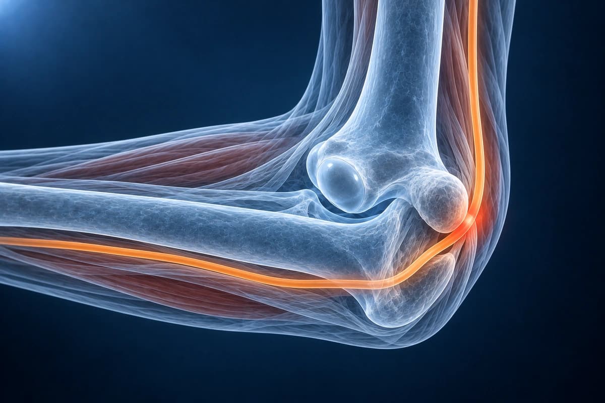
Cubital tunnel syndrome is the second most common peripheral nerve entrapment, involving compression of the ulnar nerve at the medial elbow. The nerve passes through the cubital tunnel behind the medial epicondyle.
Common Symptoms
- Numbness and tingling in the ring and little fingers that worsens with prolonged elbow flexion
- Weakness of intrinsic hand muscles causing difficulty with grip and pinch tasks
- Clawing of the ring and little fingers due to intrinsic muscle weakness in advanced cases
- Pain along the inner elbow that may radiate proximally into the arm or distally into the forearm
- Positive Tinel sign: tapping over the cubital tunnel at the medial elbow reproduces electric shock sensation
- Symptoms aggravated by sleeping with the elbow fully flexed or prolonged use of mobile devices
Key Imaging Findings
- Ulnar nerve enlargement proximal to or within the cubital tunnel
- Increased T2 signal within the nerve (neuritis)
- Nerve subluxation anterior to the medial epicondyle on flexion
- Medial epicondyle osteophytes narrowing the tunnel
- Anconeus epitrochlearis muscle (accessory muscle compressing nerve)
- Ganglion cyst or other space-occupying lesion
Frequently Asked Questions
Where exactly is the ulnar nerve compressed in cubital tunnel syndrome?
The ulnar nerve passes through the cubital tunnel at the medial elbow, a fibro-osseous channel bordered by the medial epicondyle anteriorly, the olecranon posteriorly, and the arcuate ligament of Osborne (cubital tunnel retinaculum) superiorly. The nerve is most vulnerable at the level of Osborne's ligament, where the canal narrows during elbow flexion. Additional sites of compression along the ulnar nerve course include the arcade of Struthers proximally and the flexor carpi ulnaris aponeurosis distally. Electrodiagnostic studies localizing conduction block to the elbow segment confirm the diagnosis.
How does cubital tunnel syndrome differ from ulnar tunnel syndrome at the wrist?
Cubital tunnel syndrome compresses the ulnar nerve at the elbow and affects both the dorsal cutaneous branch (supplying sensation to the dorsum of the little finger and ulnar ring finger) and the motor branch to the flexor carpi ulnaris, whereas ulnar tunnel syndrome at Guyon's canal at the wrist spares the dorsal cutaneous branch and the flexor carpi ulnaris. Sensory loss on the dorsal ulnar hand therefore points to an elbow-level lesion, while preserved dorsal hand sensation with intrinsic weakness and little finger sensory loss suggests a wrist-level compression, which may be caused by a ganglion cyst, hook of hamate fracture, or ulnar artery thrombosis.
What are the surgical options for cubital tunnel syndrome?
Simple in-situ decompression releases Osborne's ligament and the surrounding fascial constraints without moving the nerve, making it effective for mild-to-moderate disease with the advantage of lower complication rates. Medial epicondylectomy removes the medial epicondyle to eliminate the pulley effect on the nerve during elbow flexion. Anterior transposition relocates the nerve anterior to the medial epicondyle in a subcutaneous, intramuscular, or submuscular plane, reducing tension on the nerve during flexion; submuscular transposition is preferred for cases with nerve subluxation or recurrence after prior decompression. All techniques show comparable outcomes in mild disease, but severe cases with fixed intrinsic weakness have a guarded prognosis regardless of technique.
Sources
- Palmer BA, Hughes TB. Cubital tunnel syndrome. J Hand Surg Am. 2010;35(1):153-163.
- Zlowodzki M, Chan S, Bhandari M, et al. Anterior transposition compared with simple decompression for treatment of cubital tunnel syndrome. A meta-analysis of randomized, controlled trials. J Bone Joint Surg Am. 2007;89(12):2591-2598.
- Andreisek G, Crook DW, Burg D, et al. Peripheral neuropathies of the median, radial, and ulnar nerves: MR imaging features. Radiographics. 2006;26(5):1267-1287.
- Kim S, Chung BM, Kim WT, et al. Diagnosing ulnar neuropathy at the elbow on MRI: importance of the longitudinal extent of the hyperintense ulnar nerve. Skeletal Radiol. 2022;51(7):1473-1481.
Understand your imaging before your next appointment
MRI, X-ray or CT from a CD, folder or ZIP. A PDF report or photos of the films work too.
Free to open and preview, no account needed. An optional AI report from $10 explains your images in plain language, with questions for your doctor.
Your raw files stay on this device. Only rendered images and minimal context are sent when you request an AI report.
Medical Disclaimer: This page is for informational and educational purposes only. It does not constitute medical advice, diagnosis, or treatment. AI-generated analysis may contain errors. Always consult a qualified healthcare professional for medical decisions. Full Disclaimer